Welcome
Hello and Welcome to Our Home Page. learn more »
LOGIN
WHO's ON LINE
We have 499 guests and no members online
JobGrok
JG Listings
Carrier Oils
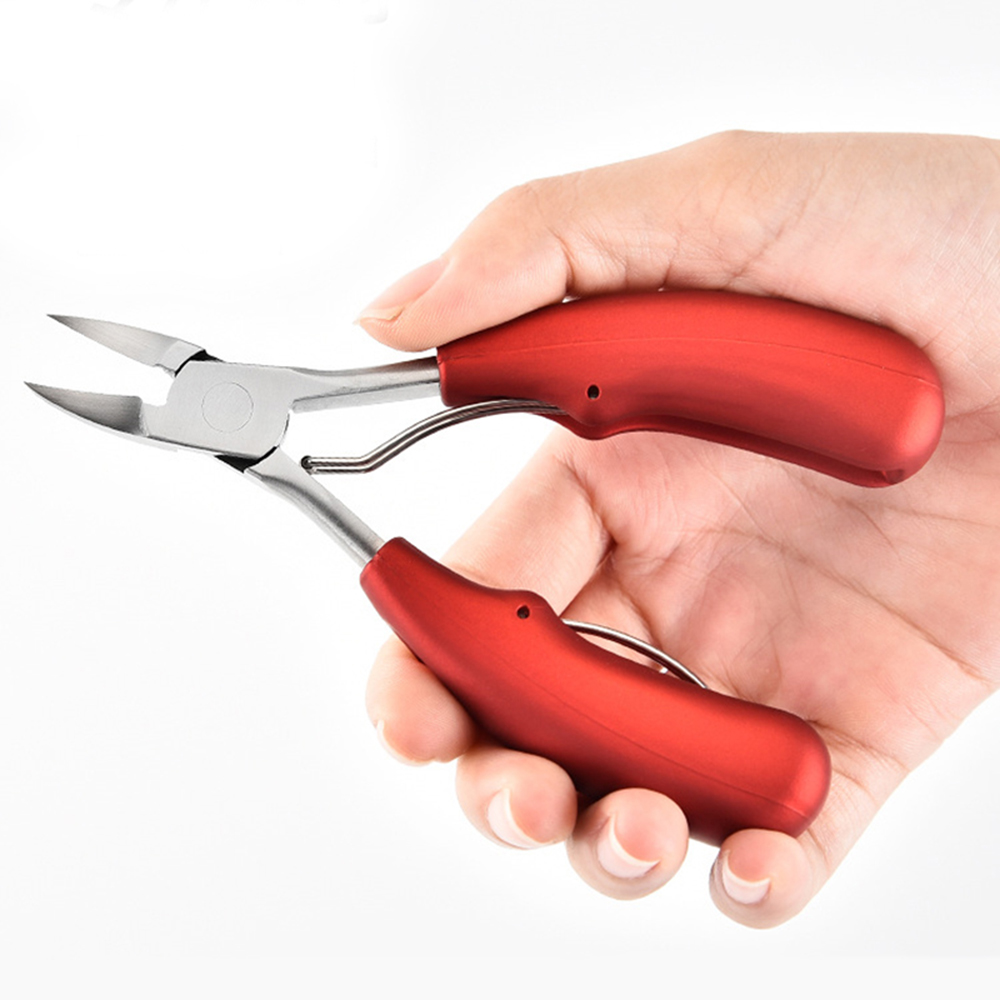
Nail Clippers
Our therapists

Ad Agency Remote
Essential Oils

Foot Structure

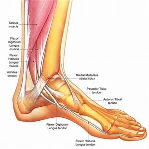
Foot Structure
MAY 25, 2018 BY CINDY SCHMIDLER 7 COMMENTS
The foot is incredibly complex. This introduction to the anatomy of the foot and ankle will be very general and highlight the main structures.
Bones and Joints of the Foot and Ankle
Ankle
The bones of the foot and ankle begin with the ankle joint itself. The ankle joint, talocrural joint, is formed where the distal end of the leg meets the foot. The ankle joint is formed where the talus (the uppermost bone in the foot) and the tibia (shin) meet. The ankle joint is both a synovial joint and hinge joint and is formed where the top of the talus fits in between the lower ends of the tibia (shin) and fibula. The talus fits between the two bony prominences on either side of the ankle. In a hinge joint, a convex part of one bone fits into the concave part of the other bone.
The two bones of the lower leg, the large tibia and the smaller fibula come together at the ankle joint to form a very stable mortise and tenon type joint. The mortise and tenon construct is well known to carpenters and craftsmen who use this type of construction to join everything from furniture to large buildings.
The ankle joint allows the foot to bend up (flex) and down (extend). When the foot is extended, the heel is drawn up and the toes point downward. The ankle joint is more stable when it is extended than when it’s flexed (toes up). Other movements, such as tilting and rotation take place in and around the joints in the foot itself.
Strong ligaments on either each side of the joint limit movement. The ankle bones are connected by the anterior talofibular ligament, posterior talofibular ligament, and calcaneofibular ligament. The articular capsule surrounds the joint with thin fibrous tissue. A synovial membrane is in under the ligaments. Of the ligaments, the deltoid is so strong that it usually resists a force which fractures the bone to which it is attached.The arteries supplying the ankle joint are derived from the malleolar branches of the anterior tibial and the peroneal.The nerves of the ankle are derived from the deep peroneal and tibial nerves.
Foot
The foot forms a firm basis of support for the body in the erect posture, and is solidly built up and its parts are less movable on each other than those of the hand. The phalanges (toes) of the foot are smaller and their movements are more limited than the fingers of the hand. Very much more different between the metacarpal bone of the thumb and the metatarsal bone of the great toe.
The foot forms a firm basis of support for the body in the erect posture, and is solidly built up and its parts are less movable on each other than those of the hand. The phalanges (toes) of the foot are smaller and their movements are more limited than the fingers of the hand. Very much more different between the metacarpal bone of the thumb and the metatarsal bone of the great toe.
The metatarsal bone of the great toe helps support the weight of the body, is constructed with great solidity, lies parallel with the other metatarsals, and has a very limited amount of movement. The tarsus forms a considerable part of the foot, and is at right angles to the leg, and relates to an erect posture. In order to support our body weight with the least amount of surface area, the tarsus and a part of the metatarsus is made up of a series of arches. The bones of the foot include the tarsals, metatarsals and phalanges.
The two bones that make up the hindfoot are the talus and the calcaneus (heel). Where the talus meets the calcaneus forms the subtalar joint. The subtalar joint allows the foot to rock from side-to-side.
Tarsal Bones
The tarsal bones work together as a group. The way these bones fit together is very interesting. When the muscles of the foot and leg twists the foot in one direction, the tarsal bones lock together and form a very rigid structure. When they twist in the opposite direction, the bones unlock, allowing the foot to conform to whatever surface the foot is contacting.
The tarsal bones work together as a group. The way these bones fit together is very interesting. When the muscles of the foot and leg twists the foot in one direction, the tarsal bones lock together and form a very rigid structure. When they twist in the opposite direction, the bones unlock, allowing the foot to conform to whatever surface the foot is contacting.
There are seven tarsal bones: the calcaneus, talus, cuboid, navicular, and the first, second, and third cuneiforms.
The calcaneus (heel bone) is the largest of the tarsal bones. It transmits the weight of the body to the ground and acts as a lever for the muscles of the calf. The calcaneus joins with the talus and cuboid.
The talus (ankle bone) is the second largest of the tarsal bones and sits atop the calcaneus. It joins on either side with the malleoli and in front with the navicular. The talus forms a joint with four bones: tibia, fibula, calcaneus, and navicular.
The cuboid is on the lateral side (opposite the arch of the foot) and in front of the calcaneus. The cuboid forms a joint with four bones: the calcaneus, third cuneiform, and fourth and fifth metatarsals; occasionally with a fifth, the navicular.
The navicular sits at the medial side of the tarsus, between the talus behind and the cuneiform bones in front. The navicular forms joints with four bones: the talus and the three cuneiforms; occasionally with a fifth, the cuboid.
The first cuneiform forms a joint with four bones: the navicular, second cuneiform, and first and second metatarsals. The second cuneiform forms joints with four bones: the navicular, first and third cuneiforms, and second metatarsal. The third cuneiform forms joints with six bones: the navicular, second cuneiform, cuboid, and second, third, and fourth metatarsals.
Metatarsal Bones
The metatarsus consists of 5 bones and are numbered starting from the arch side of the foot. The tarsal bones are connected to the 5 long bones of the foot called the metatarsals.
There is a fairly rigid connection between the two groups without much movement at the joints. The base of each metatarsal bone forms a joint with one or more of the tarsal bones, and the head with one of the first row of phalanges. The first metatarsal forms a joint with the first cuneiform, the second with all three cuneiforms, the third with the third cuneiform, the fourth with the third cuneiform and the cuboid, and the fifth with the cuboid.
Phalanges
Finally, there are the bones of the toes, called the phalanges. The joints between the metatarsals and the first phalanx is called the metatarsal phalangeal joint. These joints form the ball of the foot, and movement in these joints is very important for a normal walking pattern.
The phalanges of the foot correspond, in number and general arrangement, with those of the hand; there are two in the great toe, and three in each of the other toes. They differ from the hand, however, in their size, the bodies are shorter in length, and, especially in the first row, wider.
Not much motion occurs at the joints between the bones of the toes (phalanges). The big toe, or hallux is the most important toe for walking, and the first metatasal phalangeal joint is a common area for problems in the foot.
In the second, third, fourth, and fifth toes the phalanges of the first row form joints behind with the metatarsal bones, and form joints in front with the second phalanges, which in their turn form joints with the first and third: the ungual phalanges form joints with the second.
Soft Tissues of the Foot and Ankle
The soft tissues of the foot and ankle include ligaments, tendons, fascia, nerves and blood vessels.
Ligaments
TIP: Ligaments attach bones to bones. So ligaments are usually named after the bones they connect. The name may also contain a directional description. For example, the Posterior talotibial ligament is on the posterior side (back side) of the foot and connects the talus and the tibia.
Ligaments are strong, dense, flexible bands of fibrous connective tissue. The function of ligaments is to attach bones to bones. Ligaments help give the stability of the joint. Ligaments are often named after the the bones they join together. The ankle joint is bound by the strong deltoid ligament and three lateral ligaments: the anterior talofibular ligament , the posterior talofibular ligament and the calcaneofibular ligament.
• The deltoid ligament supports the medial side of the ankle and attaches the tibia to the calcaneus and talus.
• The talofibular ligaments join the talus and the fibula and support the lateral sides (the side opposite the arch of your foot) of the ankle.
• The calcaneofibular ligament joins the calcaneus and the fibula.
Tendons
Fascia
plantar-fascia on the bottom of the foot
Common Problems in the Ankle Joint
Ankle Sprain
Ankle sprains are one of the most common of all injuries.
Reference:https://www.healthpages.org/anatomy-function/anatomy-foot-ankle/
Articles - Latest
- Supplies, Description, and Usage - Tech Nails-2
- Supplies, Description, and Usage - Tech Nails
- Exercises for Plantar Fasciitis
- Shoes, insoles and splints: Cushioning and support - Plantar fasciitis
- 10 best bum workouts and 25 bum exercises for a 🍑'ier butt
- The dos and don’ts of running when you’re over 40
- This 30-minute workout can be done from just about anywhere
- I teach stretching routines for a living — 3 exercises that strengthen your hips and open your hamstrings
- Somatic exercise has gone viral promising to lower cortisol levels, ease stress, and boost health - so, does it actually work?
- Planks and wall sits best exercise for lowering blood pressure, study says
- Four moves and six minutes is all you need to develop strength with this no-equipment routine
- I did a two-minute Farmer’s Carry every day for a week — here’s what happened
- No squats or lunges: This knee-friendly workout sculpts your lower body in 7 exercises
- Two dumbbells and five moves are all you need to build strength in the shoulders and back
- Add Muscle, Build Stamina and Fire up Your Metabolism with Our Three-Move Strongman Circuit
- How to clean running trainers without ruining them: 3 easy steps to take
- Podiatrist shares pain-inducing mistakes we're making when wearing high heels
- 9 things you need to know before getting acrylic nails
- Running could be just as effective at treating depression as medication, scientists find
- This is How Long You Should Rest Between Sets, Whatever Your Training Goal
- Manicurists Share Their Top Tips For Growing Stronger Nails Without Giving Up On Those Gel Manis
- Dry, cracked heels: Here are the most moisturizing ingredients according to a dermatologist
- Treatment for Plantar Fasciitis
- How to measure your feet: A guide for runners
- Heel and ankle conditions and Injuries
Articles-Popular
- Home
- Calluses and Corns-4-Padding and Insoles To relieve Pressure
- Contacts
- Appreciate Your Feet
- WEB - LINKS
- Therapy Price List- Aromatherapy - Counselling
- The Awareness of Foot Care
- Nail Technician Resume
- Join us as a Therapist
- Gallery - Pedicured Feet
- Podiatry/Chiropody Price List
- Skin Care-Feet
- TCM - Therapy Prices
- Blisters on the Feet
- Nail Technician Job Description
- Galleries
- Bacterial Infections
- Itching Skin on the Feet
- Athlete's Foot
- Sweaty or Smelly Feet
- Appointments
- Skin Changes Associated with Blood Flow
- Common Toenail Conditions - Changes in Nail Colour
- Calluses and Corns - 2
- Insoles
